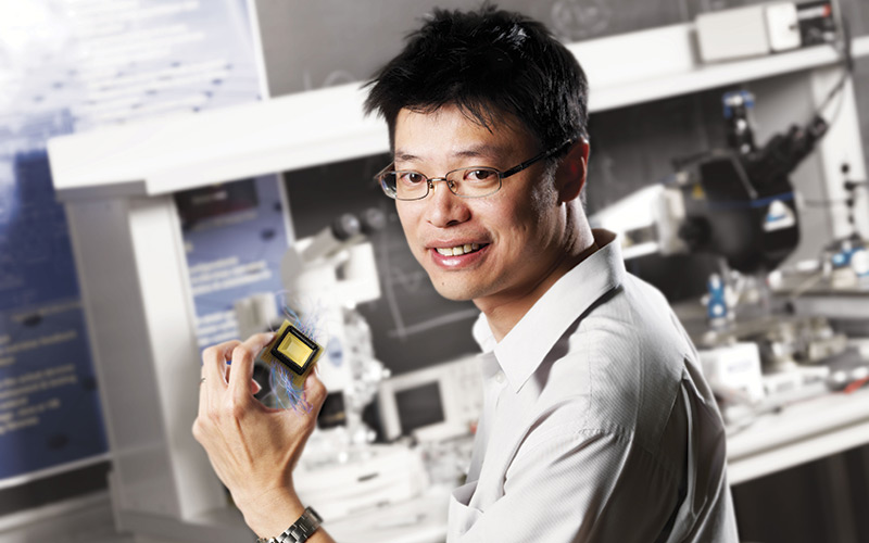
A microscope that uses artificial intelligence to develop 3D images has been created.

The second-generation microscope can produce large, full-colour images of tissue and fluid samples.
It is expected to be available at a cost of hundreds of pounds – potentially reducing costs for disease diagnosis in developing countries.
The spectral light-fusion microscope has no lens, but uses artificial intelligence and mathematical models of light to construct an image. Alexander Wong, the Canada Research Chair in Medical Imaging at the University of Waterloo, co-led the research project.
He said: “We know that pathology is the gold standard in helping to analyse and diagnose patients, but that standard is difficult to come by in areas that can’t afford it.
“This technology has the potential to make pathology labs more affordable for communities who currently don’t have access to conventional equipment.” Details of the first-generation microscope were published last year.
More information can be found here.


