Barbara J. Bain, Professor of Diagnostic Haematology and Honorary Consultant Haematologist, analyses spherocytes and irregularly contracted cells.
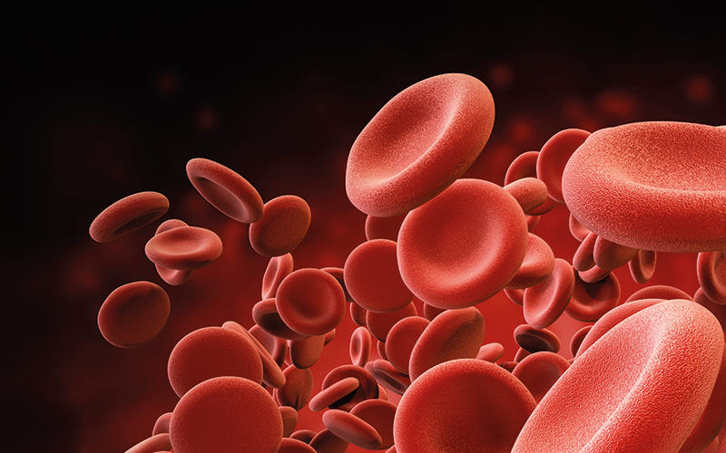
Spherocytes and irregularly contracted cells have morphological similarities but also differences. Their clinical significance is quite different, so it is important to make a distinction. Both cell types have reduced central pallor indicating that the normal disciform shape has been lost. However, spherocytes have a perfectly circular outline (Fig. 1, top right) whereas irregularly contracted cells have an irregular outline (Fig. 2, middle right).
Spherocyte formation
Spherocytosis results from an inherited or an acquired abnormality of the red cell membrane. Spherocytes are formed when there is loss of red cell membrane with the cell volume being fairly well preserved. A distinction should be drawn between spherocytes and microspherocytes, the latter resulting from red cell fragmentation in which there is loss of cell volume but with a disproportionate loss of membrane.
They are also sometimes known as spheroschistocytes. Causes of both are shown in Table 1 (below). Hereditary spherocytosis can result from a mutation in a variety of genes encoding ankyrin (ANK1), β spectrin (SPTB), the red cell membrane protein designated band 3 (SLC4A1), the α spectrin gene (SPTA) or the protein 4.2 (palladin) gene (EPB42). As the result of the mutation, there is reduced synthesis of the protein in question. This leads to loss of the normal tethering of the lipid bilayer to the underlying cytoskeleton so that pieces of the membrane become detached. In the immune causes of spherocytosis (autoimmune, alloimmune and drug-induced immune) the mechanism is recognition by splenic macrophages of immunoglobulin or complement components bound to the red cell membrane with resultant removal of some of the membrane by the macrophage.
Please click here to read the full article.
Image credit |Shutterstock