Biomedical Scientist Team Manager Tahmina Hussain puts a new automated digital analyser to the test.
Full blood count samples requiring morphological examinations are one of the most valuable investigations in the haematology laboratory, due to their significant contribution towards the diagnosis and monitoring of disease processes. It is imperative that these procedures can be completed as quickly as possible, especially in cases where immediate clinical action is required.
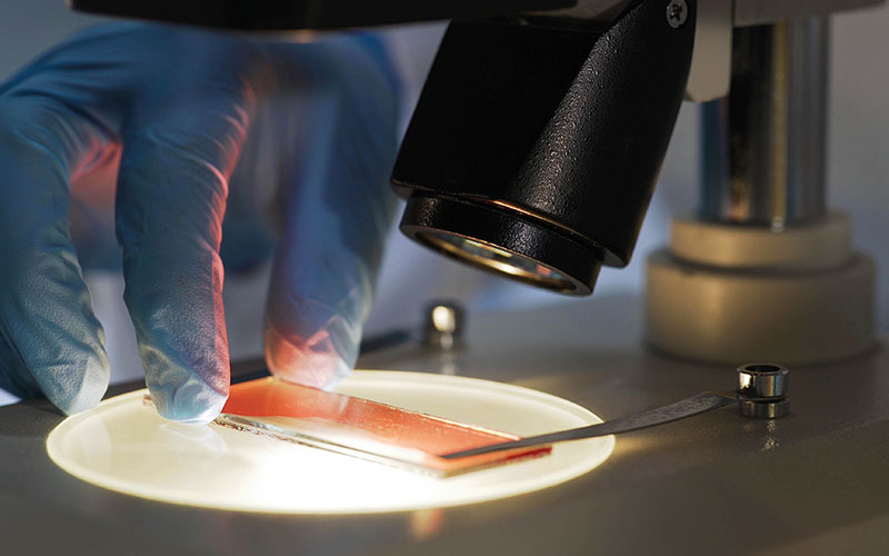
Manual light microscopy is considered the gold standard for reporting morphology of blood cells. This method is time consuming at The Christie NHS Foundation Trust, where the maindiagnoses in the haematology laboratory consist of haematological malignancies, and the presence of abnormal white blood cells contributes towards the heavy workload. Limitations include subjective interpretation of cells and variation in results reported between individuals. Due to the nature of the malignancies being monitored post-chemotherapy, many samples are leucopenic, which carries a further challenge of counting and identifying the adequate number of cells required in order to provide an accurate manual differential. Development of automated digital morphology analysers has made it possible to identify white blood cells, which improves turnaround times and reduces subjectivity in the interpretation of cells.
Digital morphology
Performing manual differentials is highly demanding on time, as well as requiring experienced staff and this has an effect on turnaround times, particularly during night shifts and weekends, when the availability of trained staff may be limited. The introduction of a digital morphology analyser may be a solution to overcoming these challenges. Additional benefits of introducing a digital morphology analyser in the haematology laboratory include speeding up the process for training on morphology, as the database stores images of the cells examined in the peripheral blood smears in a reference library that is accessible to trainees. The communication between staff and consultants can be improved as the blood smears can be reviewed through email or by logging into the software system remotely.
Please click here to read the full article
Please click here to read all the Science articles from The Biomedical Scientist