A recent advance in histopathology eliminates the need for chemical staining and enables high-resolution imaging of thick tissue sections.
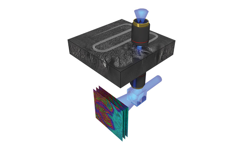
An international research team demonstrated a 3D label-free quantitative phase imaging technique that uses optical diffraction tomography to obtain volumetric imaging information. Automated stitching simplifies the image acquisition and analysis.
The new optical diffraction tomography method enables label-free high-contrast visualisation of transparent samples.
The team used digital refocusing and automated stitching, enabling volumetric imaging of 100 µm-thick-thick tissues over a lateral field of view of 2 mm by 1.75 mm while maintaining a high resolution of 170 × 170 × 1400 nm. They demonstrated that simultaneous visualisation of subcellular and mesoscopic structures in different tissues is enabled.
Image credit | Hugonnet et al